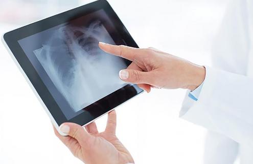
Patients who come to a Dartmouth-Hitchcock Health (D-HH) facility for a diagnostic exam requiring an X-ray will now notice their technologist no longer draping a heavy lead apron, also known as gonadal shielding, over their lap. It is now widely recognized that X-ray imaging is safer and more effective by limiting the use of the traditional practice of routine lead apron shielding during an X-ray. This shift in guidelines was first championed by the American Association of Physicists in Medicine and the American College of Radiology and has now been instituted at D-HH locations.
“Studies have shown that shielding patients provides very little to no benefit. When a patient receives an X-ray, we take care to only irradiate the area necessary for the exam,” said Michael Timmerman, Radiation Safety Officer at Dartmouth-Hitchcock Medical Center (DHMC). “Lead only protects you from radiation outside of your body. When you receive an X-ray, a small amount of radiation will get to other areas of your body, but this occurs internally; lead will do nothing to block this radiation.”
There had been previous concerns that radiation might damage cells that could be passed along to future generations, however, these concerns have not been proven by any medical studies to date. Because of this concern, lead shields were often placed over patients’ reproductive organs during medical imaging exams. Since the 1950s, the amount of radiation used in medical imaging has decreased over 95 percent, and advances in imaging equipment now can produce quality images using small amounts of radiation. According to the American Association of Physicists in Medicine, the presence of the lead apron can also impair the quality of diagnostic tests.
“D-HH is committed to keeping radiation doses to our patients as low as possible. Therefore, we keep our equipment up-to-date and continue to invest in newer X-ray technologies that reduce radiation per image,” said Timmerman. “Our technologists and physicians are trained to use the smallest amount of radiation necessary for the exam. We also consider using non-radiation options (such as ultrasound or MRI) when appropriate.”